bodybuilding & Fitness
The anatomy of the back muscles and the correct method for targeting each muscle are discussed.
Muscle anatomy is essential for all bodybuilders to attain muscle mass and strength. As a result, it is critical to investigate and understand the muscle anatomy of each back region.
We classify the back muscles as drawing muscles, indicating that they contract when the body pulls them inward.
Focusing on the back muscle necessitates a scientific understanding of its internal workings, given its status as one of the body’s most significant muscles. Consequently, it is imperative to investigate the anatomy of the back muscles.
Given that these muscles are present in four of the seven display positions, it is imperative to train them with a high level of concentration, increase the training load, and incorporate a variety of exercise methods to maximize their potential.
Additionally, in order to develop substantial back muscles, it is essential to comprehend the anatomy of the back muscles. This knowledge will enable you to identify the most effective exercises for beginners.
The most crucial elements within the anatomy of the back muscles
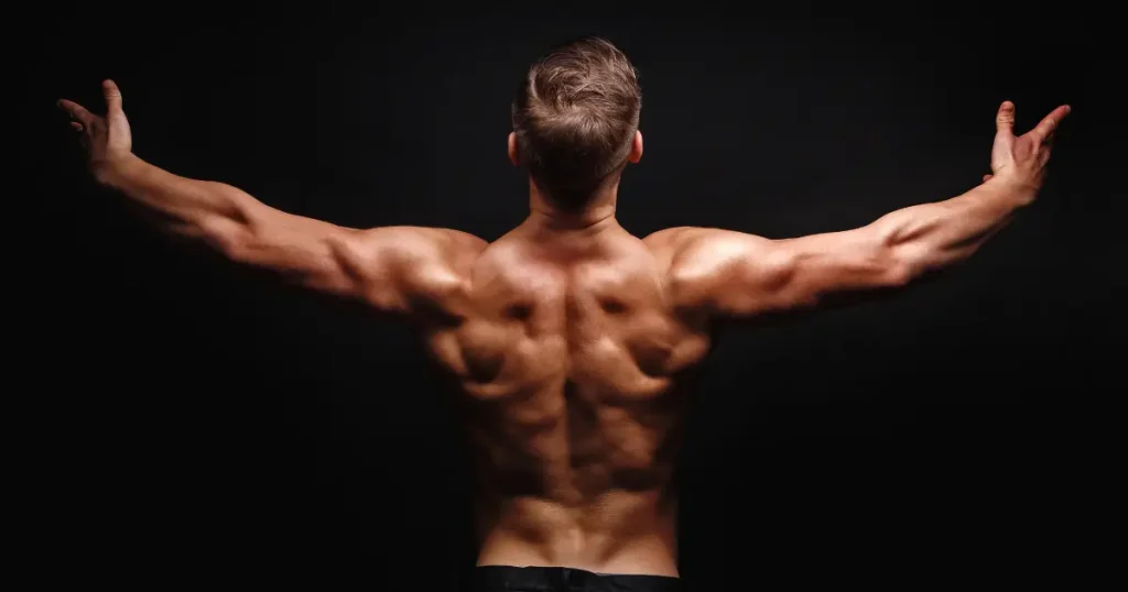
Prior to commencing the dissection of the back muscles, it is important to note that they are the second largest muscle in the body, following the leg muscles. Consequently, they contribute to the player’s most aesthetically pleasing appearance, which is the (v) shape. Furthermore, they are responsible for the body’s wider appearance than the shoulders. In this article, we will elucidate the anatomy of the back muscles to enable you to target each muscle in the appropriate manner.
Arabic terms for the back muscles
- Lateral or lateral back muscle
- Trapezius muscle
- The rhomboid, or middle back, muscle
- Lumbar or lower back muscle
- Rotator cuff tissue
1. Lateral or lateral back muscle: Latissimus dorsi.
The arm bone, one of the spine’s bones, regulates the lateral back muscle. Moving the arm vertically activates and extends the lateral back muscle, resulting in its maximum extension while the spine remains stationary.
Therefore, to ensure adequate activation of the muscle, apply a pull from above. The lateral back muscles, known as the “Lats” in English, primarily perform this exercise, which we refer to as the lat-pulldown or the wide front pull.
The lateral back muscle is the largest component of the back muscle and is responsible for the V shape or wings that the back muscle possesses. Therefore, you should use any exercise that involves a vertical pull to activate the lateral back muscle. Through this explanation, you now understand the back muscles in this area.
What are the most effective exercises for the lats muscle?
The trapezius muscle
The dissection of the back muscles revealed that the upper back muscle consists of three distinct components: an upper, middle, and lower part. Furthermore, it maintains a stationary connection with the shoulder blade and the clavicle.
Because the muscle connects to the shoulder blade, it’s crucial to keep the shoulder blade unobstructed. Avoid closing the shoulder blade, which limits range of motion and reduces exercise benefits.
Therefore, in order to achieve the most effective activation of the upper back muscle, it is necessary to pull from the front to the back. The most effective method of lifting the weights is horizontally.
By maintaining an inclined position with your body, you will provide the shoulder blade with the maximum extension, resulting in the most activation.
Therefore, perform the exercise while seated on a chair at a 45-degree angle with your chest supported, and pull the dumbbell up to achieve the maximum activation of the upper back muscle.
Therefore, the upper back exercise, also known as “dumbbell shrugs” or “shaking the bolt with dumbbells,” is not the most efficient way to activate the muscle. However, it is a beneficial exercise.
This is because the exercise is horizontal and involves a pull with the shoulder blade movement. Unquestionably, contraction highly activates the muscle, but it’s the extension and negative movement, involving descending, that actually build the muscle.
3. The muscle known as the rhomboid or medial back
Two superficial muscles located in the upper part of the back compose the rhomboid muscle. We refer to these muscles as the rhomboid minor and the rhomboid major due to their functional similarity. Together with the posterior shoulder and the latissimus dorsi muscle, or lats, the rhomboid muscle forms the superficial layer of the external back muscles.
Between the thoracic vertebrae and the shoulder, the rhomboid muscles are responsible for medially retracting the scapula. As a result, the rhomboid is critical for stabilizing the scapula in its proper position and strengthening the shoulder.
The primary function of the rhomboid muscles is to retract the shoulders around the posterior shoulder joint.
Shoulder retraction is the movement of the shoulder bone upward and medially along the back, which rotates the shoulder downward. By significantly extending the shoulder joint, the rhomboid will help you maintain the correct position while sitting, standing, and walking.
Furthermore, the rhomboid functions to secure and stabilize the scapula in its current position. This serves as a fulcrum around which the upper body can move as well as a point of attachment between the shoulder and the back, allowing the various upper body muscles to function.
Therefore, we can infer that the rhomboid muscle rests beneath the trapezius muscle, firmly attached to the scapula. The rhomboid muscle is able to move in response to the scapula’s movement.
It is crucial to avoid closing the scapula during these exercises, as this will limit the range of motion and hinder our ability to derive the maximum benefit from the exercises. When moving the scapula, draw horizontally.
4. Lumbar or Lower Back Muscle
The term “spinal” specifically refers to the vertebrae in the back. The term “spinal” refers to the back vertebra. After dissecting the back muscles, it is clear that the spinal muscles are a group of muscles located along the spine. The rotators and multifidus muscles are additional muscles in the muscle group that surrounds the spine.
In general, the spinal muscles are also present.
The erector spinae are the closest to the spine because they are located next to the lumbar region, which is where the spinal muscles are primarily located in the midsection.
In the lower back, the diamond-shaped lumbar muscle surrounds the spine or vertebral column. The lumbar muscle is considered the most crucial muscle in the back region, serving as the foundation for both the back and the entire body. Without the lumbar, the body is akin to a building without a foundation. Consequently, it is impossible to construct a strong back without a strong lumbar. You can concentrate on this muscle by performing exercises that engage the entire upper body.
5. Rotator tendon
Four muscles and their associated tendons comprise the rotator cuff. Each of them contributes to your shoulder’s specific movement, and when combined, they stabilize your back shoulder and maintain the position of your upper arm.
All four rotator cuff muscles originate from your shoulder blade, but their other extremity connects to different parts of your upper arm bone.
The symptoms of rotator cuff injuries vary depending on the structure of the back muscles and may include:
- A dull ache is typically characterized as pain in the shoulder area.
- It can be challenging to extend your arm during daily activities, such as raising your palms.
- Shoulder muscle weakness or rigidity.
- The pain intensifies at night, obstructing sleep on the affected side.
- When you move your arm, you may hear popping or crackling noises.
However, some individuals who have sustained a rotator cuff injury may not experience any discomfort. The condition may progress and deteriorate over time. Only approximately one-third of rotator cuff injuries result in pain.